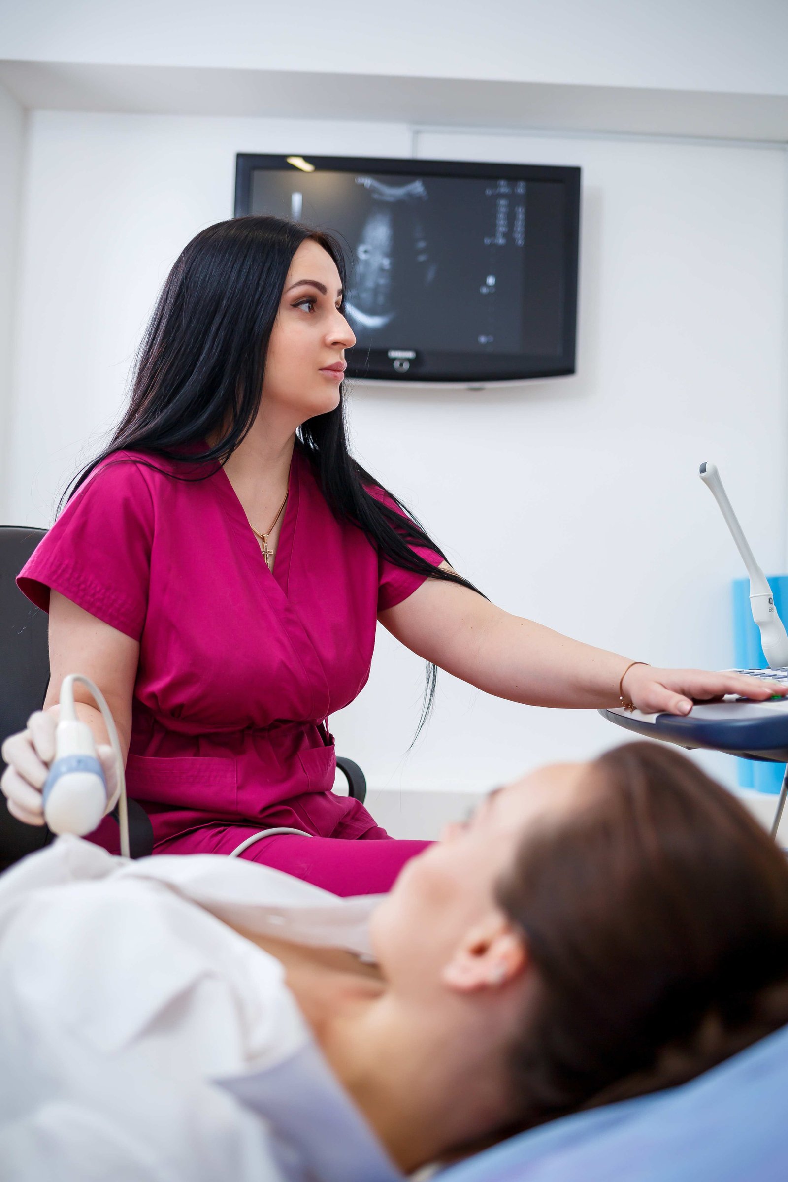
2D Echocardiography Cardiac Ultrasound Setup

2D Echocardiography (2D-Echo) & Colour Doppler

The Department of Non-Invasive Cardiac Diagnostics at Aditya Hospital (Aditya Lifeline Pvt. Ltd.) provides high-resolution 2D Echocardiography (2D-Echo) with Colour Doppler imaging in Indore. 2D-Echo is a completely non-invasive, radiation-free ultrasound investigation that produces real-time cross-sectional images of the beating heart, enabling our cardiologists to accurately assess cardiac structure, chamber dimensions, myocardial wall motion, and valvular function.

1. Clinical Importance & Diagnostic Capabilities

2D-Echo is the gold standard primary diagnostic tool for evaluating a wide range of cardiovascular conditions and guiding appropriate medical or surgical therapies:

  • Ventricular Function & Ejection Fraction (EF): Precise quantification of systolic and diastolic pumping performance of the left and right ventricles.
  • Valvular Heart Disease Assessment: Detailed detection of stenosis (narrowing), regurgitation (leakage), and prolapse in mitral, aortic, tricuspid, and pulmonary valves.
  • Wall Motion Abnormality (RWMA): Early identification of localized myocardial hypokinesia or akinesia indicative of previous or evolving myocardial infarction (heart attack).
  • Pericardial & Congenital Anomalies: Fast detection of pericardial effusion (fluid around the heart), cardiac tamponade, atrial/ventricular septal defects (ASD/VSD), and intracardiac thrombi/clots.

2. When Is a 2D-Echo Recommended?

Physicians and cardiologists frequently recommend a 2D Echocardiogram for patients experiencing:

Chest Pain & Shortness of Breath

Evaluation of unexplained dyspnea (breathlessness), exertion fatigue, angina pain, and suspected congestive heart failure.

Heart Murmurs & Valvular Clicks

Investigating abnormal heart sounds or murmurs detected during routine clinical auscultation to rule out valve pathology.

Pre-Operative Cardiac Fitness

Essential pre-anesthetic check (PAC) clearance to evaluate cardiovascular safety prior to major general or laparoscopic surgeries.

Chronic Hypertension & Diabetes

Monitoring long-term hypertensive changes such as left ventricular hypertrophy (LVH) and diabetic cardiomyopathy.

  • Completely Painless, Safe, and Zero-Radiation Ultrasound Imaging
  • High-Definition Colour Doppler & Continuous / Pulsed Wave Doppler
  • Immediate Diagnostic Reports Reviewed by Senior Cardiologists
  • Bedside Portable 2D-Echo Available for ICU & Critical Inpatients

Diagnostic Expertise & Specialist Team

Our cardiac diagnostic suite is led by experienced cardiologists and sonologists ensuring detailed imaging interpretation and prompt reporting:

CONSULTANT CARDIOLOGIST
MD, DM (Cardiology) Echo & Clinical Panel
CARDIAC SONOLOGIST
Echocardiography Specialist Doppler Diagnostics
CRITICAL CARE UNIT
Intensive Care Team Bedside ICU Echo

3. How the Procedure is Performed & Patient Preparation

A standard transthoracic 2D Echocardiogram is quick, straightforward, and requires virtually no strenuous preparation:

  • No Fasting Required: Patients can eat, drink, and take their routine prescribed medications normally prior to the test.
  • Procedure Duration: The scan typically takes between 15 to 25 minutes in a relaxed, dimmed-light examination room.
  • Comfortable Positioning: The patient lies on their left side while water-soluble acoustic gel is applied to the chest to obtain clear sonic windows.
  • Immediate Activity Resumption: There is zero recovery downtime or sedative effect; patients can drive home or resume work immediately after the test.

4. Integrated Non-Invasive Cardiac Support

2D-Echo works seamlessly alongside our 12-lead ECG, Treadmill Stress Testing (TMT), and pathology cardiac biomarker profiling, ensuring an exhaustive, 360-degree cardiac workup under one roof at Aditya Hospital.

Book Your 2D-Echo Test Today

Appointments and walk-in diagnostic investigations available at Aditya Hospital, 318, Usha Nagar Ext., Indore.

Diagnostic Desk:
+91 0731 2483311